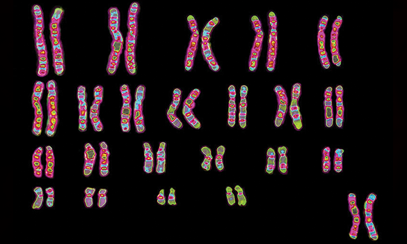
What is a Karyotype?
Imagine your body as a beautifully complex puzzle, with each piece playing a crucial role in creating who you are. This puzzle is your genetic blueprint, written in the language of DNA and organized into structures called chromosomes. But how do we make sure everything is in the right place and functioning correctly? That’s where karyotyping comes in.
Karyotyping is like taking a snapshot of your chromosomes, the tiny thread-like structures inside your cells that carry your genetic information. To get this snapshot, doctors use a special test that can be done in a few different ways.
How is a Karyotype Created?
- Sample Gathering: The first step in the process involves taking a small sample of cells from the mother. This can be done through a simple blood test or by collecting amniotic fluid from around the baby. These cells are then examined to check for any potential genetic issues.
- Cell Culture: The goal of growing these cells in the lab is to encourage them to divide. Cells are easiest to study when they're dividing because that's when the chromosomes are most visible. By making sure the cells go through division, scientists can get a clearer look at the chromosomes and better understand their structure and any potential issues.
- Harvesting Cells: When cells start dividing, doctors sometimes use drugs to pause them at a specific stage called metaphase. This is when the chromosomes are most condensed and easiest to see under a microscope. By stopping cells at this stage, scientists can better study the chromosomes and check for any problems or abnormalities.
- Imaging and Staining: Giemsa and other dyes are used to color chromosomes in a way that helps scientists see them clearly under a microscope. These dyes stick to specific parts of the DNA, creating a unique banding pattern on the chromosomes. This pattern makes it easier to identify and study the chromosomes, much like how a colorful map helps you navigate and understand different areas.
- Analysis: After taking a photo of chromosomes, scientists arrange them in pairs based on their size, banding patterns, and where their centromeres (the central parts of the chromosomes) are located. This organized arrangement is called a karyogram. It’s like putting together a photo album where each page shows a different set of chromosome pairs, helping doctors and researchers easily spot any abnormalities or issues.
What Makes Karyotyping Crucial?
Several essential purposes of karyotyping in medicine and research are as follows:
- Genetic Disorder Diagnosis: Karyotyping can be used to detect chromosomal abnormalities including Turner syndrome (monosomy X), Klinefelter syndrome (XXY), and Down syndrome (trisomy 21). These disorders are caused by modifications to the number or makeup of chromosomes.
- Diagnosis of Cancer: Chromosome abnormalities are frequently linked to cancer. Karyotyping aids in cancer diagnosis, prognosis determination, and treatment plan selection.
- Prenatal Testing: Karyotyping is a test used during pregnancy to check for genetic issues in the baby. To do this, doctors might use procedures like chorionic villus sampling (CVS) or amniocentesis.Both tests help doctors find out if the baby has certain genetic conditions, giving parents important information to plan for the future.
- Research and Understanding Evolution: Chromosomal evolution, genetic diversity, and the evolution of species have all benefited from the use of karyotyping.
Types of Chromosomal Abnormalities Detected by Karyotyping
- Numerical Abnormalities: These include chromosome abnormalities, such as those associated with Turner syndrome (monosomy X) or Down syndrome (trisomy 21).
- Structural Abnormalities: These involve changes in the structure of chromosomes, such as deletions, duplications, inversions, and translocations. For example, in chronic myeloid leukemia (CML), there's a specific swap, or translocation, between chromosomes 9 and 22. This particular change is often linked to the disease and helps doctors diagnose it.
The Drawbacks of Stereotyping
Although karyotyping is an effective technique, it has drawbacks:
- Resolution: Karyotyping is useful for spotting big changes in chromosomes, like when there's an extra or missing chromosome. However, it can’t detect smaller genetic changes. For instance, it might miss tiny mutations, small deletions, or tiny duplications in the DNA. So, while karyotyping can reveal major genetic issues, it’s not designed to catch every possible genetic problem.
- Detection Resolution: To spot tiny genetic changes, doctors use methods like array comparative genomic hybridization (aCGH) and fluorescent in situ hybridization (FISH). Think of aCGH as a very detailed map that highlights even the smallest differences in your genes, while FISH uses special colours to mark specific parts of your DNA. Both techniques offer a clearer view of your baby's genetic makeup, helping to identify issues that might not be visible with less detailed tests.
Progress Beyond Karyotyping
More complex genetic analysis techniques have been made possible by technological advancements:
- FISH (Fluorescent In Situ Hybridization): It’s a method that helps find tiny missing or extra pieces in chromosomes by looking for specific DNA patterns. Basically, it’s like using a magnifying glass to spot small changes in the genetic material, which can be important for identifying certain genetic conditions.
- Microarray Analysis: This technique provides a closer look at any problems with chromosomes, making it easier to spot changes in the DNA. This means we can catch issues more accurately and early on, helping doctors and parents understand the baby’s health better.
- Next-Generation Sequencing (NGS): While traditional karyotyping can show big changes in chromosomes, next-generation sequencing (NGS) goes much further. It looks at the genome in much greater detail, detecting even the smallest changes in individual DNA building blocks. This means NGS can find subtle genetic alterations that karyotyping might miss, giving a more complete picture of a person's genetic makeup.
Karyotyping is like taking a detailed look at a baby’s chromosomes to check for any abnormalities. By examining these chromosomes, doctors can spot issues that might affect the baby’s health.
In conclusion, understanding your baby’s chromosome patterns through karyotyping can provide valuable insights and help you make informed choices about your pregnancy. It’s all about giving you the information you need to prepare and support your family in the best way possible.