Slide 1

Care Begins Before Birth

"Competent Fetal & Maternal medical care, comprehension, and administration. Your well-being is our first concern! Empowering your health journey with expert attention."

Latur Fetal Medicine Centre

Established in 2022, Latur Fetal Medicine Centre is the first dedicated fetal care unit in Latur district and its suburbs. Our mission is to deliver the highest quality treatment and diagnosis for both fetal and maternal health. As a comprehensive one-stop center for mothers and babies, we provide advanced fetal medicine services using state-of-the-art technology. In today's era... Read More.

We Have Successfully Completed

Advance ultrasonography

Total Procedures

Genetic Counselling

Testimonials

Our Services

service

Advance Fetal Ultrasonography

  • NT scan (11 to 13 wk.)
  • Early Anomaly scan (16 to 18 wk.)
  • Genetic Sonogram (16wk to 18 wk.)
  • Anomaly scan (18 to 20 wk.)
  • Neurosonogram (23 wk.)
  • Echocardiography (22 wk.)
service

Genetics Study & Counselling

  • Genetic counselling online and offline
  • Downs syndrome
  • Edward syndrome
  • Patua Syndrome
  • Thalassemia
  • Sickle cell anemia
  • Single gene disorder
  • Other
service

Procedures

  • Fetal Reduction (11 to 13 wk.)
  • Chorionic villi sampling (11 to 13 wk.)
  • Amniocentesis (16wk onward)
  • Fetal Blood Transfusion
  • Amnioinfusion

Latest Blogs

Amniocentesis

Amniocentesis & CVS

October 17, 2024

Prenatal testing has revolutionized obstetrics and gynecology, offering families crucial insights into the health of their unborn child. Two of the most important practices in this field are Chorionic Villus Sampling (CVS) and Amniocentesis. These tests provide families with peace of mind and aid in decision-making by identifying genetic and chromosomal disorders.


What is Amniocentesis?

Amniocentesis is a prenatal test performed between weeks 15 and 20 of pregnancy. During this procedure, a small amount of amniotic fluid, which surrounds the fetus in the uterus, is carefully extracted. This fluid contains fetal cells and various proteins that can be analysed to detect genetic conditions. By examining these cells and proteins, doctors can provide valuable information about the baby’s health and development.


Procedure of Amniocentesis:


  1. Preparation: To choose a safe location for the needle insertion and to ascertain the position of the fetus, the procedure starts with an ultrasound.


  1. Extraction: To remove a little sample of amniotic fluid from the mother, a thin needle is delicately pushed through the mother's abdominal wall and into the uterus.


  1. Analysis: The fluid is subsequently transported to a lab for in-depth examination, where fetal cells are grown and checked for genetic anomalies.


Why is Amniocentesis Done?

Generally speaking, women who are more likely to give birth to a child who has a genetic problem should consider amniocentesis. Women over 35, those with a family history of genetic illnesses, and those whose prenatal test results have been abnormal are also included in this. It is capable of identifying ailments including spina bifida, cystic fibrosis, Down syndrome, and more.


Benefits and Risks:

Amniocentesis is a valuable diagnostic tool that provides highly reliable results for various conditions, but it does come with some risks. Because it's an invasive procedure, there’s a small chance of infection or miscarriage. It's important for parents to have a thorough discussion with their healthcare provider about these potential risks and benefits before deciding whether to proceed. This conversation can help ensure that they make an informed choice that aligns with their comfort levels and health goals.


What is Chorionic Villus Sampling (CVS)?

Another prenatal test that is carried out before amniocentesis is called Chorionic Villus Sampling, or CVS. It is usually carried out between the tenth and thirteenth weeks of pregnancy. Using CVS, a tiny sample of placental tissue which has the same genetic material as the fetus, allowing doctors to check for genetic conditions and other issues early on. This test offers vital information earlier in the pregnancy and can diagnose many of the same issues as amniocentesis.


Procedures of CVS:


  1. Preparation: To find the fetus and placenta, an ultrasound is performed initially, the same as in amniocentesis.


  1. Extraction: A tiny tube or needle is used to take a sample through the cervix (transcervical) or the abdominal wall (transabdominal), depending on where the placenta is positioned.


  1. Analysis: To find any genetic anomalies, a lab analyses the placental tissue sample.


Why is CVS Done?

CVS, like amniocentesis, is advised for women who are more likely to give birth to a child who has a genetic problem. It is capable of identifying diseases including sickle cell anemia, cystic fibrosis, Down syndrome, and other genetic abnormalities. Compared to amniocentesis, CVS has the benefit of early discovery, which enables quicker decision-making and, if necessary, intervention.


Benefits and Risks:

CVS offers the advantage of early implementation, which provides parents with more time to explore their options. However, like amniocentesis, CVS is an invasive procedure that carries risks, such as infection, miscarriage, and, if performed too early, potential limb abnormalities. Given these concerns, it’s essential for parents to have a detailed and supportive consultation with a healthcare professional. This ensures they are fully informed and can make the best decision for their situation.


Amniocentesis and Chorionic Villus Sampling (CVS) are important prenatal tests that help detect genetic conditions in a developing fetus. While both tests carry some risks, they offer valuable insights that can be crucial for early diagnosis and management of potential health issues. It's essential for parents to have open and detailed discussions with their healthcare providers to understand the benefits and risks, helping them make informed decisions that are best for their family's future.


At our clinic, we understand the importance of providing comprehensive and compassionate care for you and your baby. Whether you're considering amniocentesis or chorionic villus sampling (CVS), our specialised care team in fetal medicine is here to support you every step of the way. Our experienced professionals are dedicated to ensuring that you receive the best possible care, addressing any questions or concerns you might have with empathy and expertise. Your well-being and that of your baby are our top priorities, and we’re committed to guiding you through these important decisions with the utmost care and support.

Thalassemia & Sickle Cell Disease

Thalassemia & Sickle Cell Disease

October 9, 2024

The quality of life and general health of an individual can be greatly affected by blood diseases. Thalassemia and sickle cell disease are two examples of such hereditary blood disorders. Both disorders have an impact on the body's capacity to generate healthy red blood cells, which can result in many health issues. 


What is Thalassemia?


Thalassemia is a genetic blood disorder that affects the body's ability to produce enough haemoglobin, the protein in red blood cells responsible for carrying oxygen throughout the body. When haemoglobin levels are low, it can lead to anaemia and other complications. There are two main types of thalassemia: alpha and beta thalassemia. Each type impacts a different part of the haemoglobin molecule, leading to varying symptoms and severity.

Alpha thalassemia occurs when there is a defect in one or more of the four alpha-globin genes. The severity depends on how many of these genes are affected. Beta thalassemia, on the other hand, involves defects in one or both of the two beta-globin genes. People with beta thalassemia may experience more severe symptoms, depending on whether one or both genes are affected.


Symptoms of Thalassemia:

The kind and degree of thalassemia can affect the symptoms. Typical signs and symptoms include:

  • Weakness and exhaustion
  • Pale or tan skin
  • Abnormalities of the facial bones
  • Gradual expansion
  • Abdominal swelling


Causes of Thalassemia:

If both parents carry the gene for thalassemia, there's a higher chance their child will inherit the condition. Thalassemia is passed down from parents who both have the trait, making it more likely for their child to develop the condition.


Handling and Medical Interventions:

Regular blood transfusions to keep haemoglobin levels in check, iron chelation therapy to eliminate extra iron from the body, and occasionally a bone marrow transplant are all part of the treatment for thalassemia. Effective management of Thalassemia necessitates an early diagnosis and continued medical attention.


Sickle Cell Disease: What Is It? 


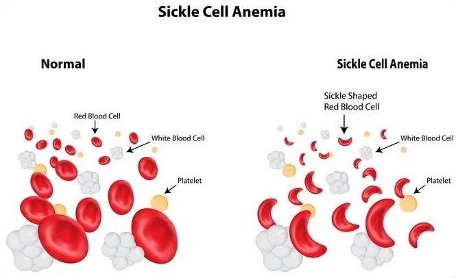
Sickle cell disease (SCD) is a genetic blood disorder where red blood cells become rigid and shaped like crescents or sickles instead of the usual round shape. These misshapen cells can block blood flow in small blood vessels, which can lead to pain, organ damage, and other serious health issues. The obstructed blood flow can cause severe pain episodes, known as crises, and may result in damage to organs like the spleen, liver, and kidneys.


Symptoms of Sickle Cell Disease:

People who have sickle cell disease may encounter:


  • Painful episodes(sickle cell crises)
  • Anaemia and exhaustion
  • Edema in the feet and hands
  • Recurring infections
  • Children's delayed growth 


Causes of Sickle Cell Disease:

Because sickle cell disease is inherited in an autosomal recessive manner, meaning a child must receive the sickle cell gene from both parents to have the disease. If both parents carry the gene, there's a 25% chance with each pregnancy that the child will have sickle cell disease. People of African, Mediterranean, Middle Eastern, and Indian origin are the most commonly affected by the illness.


Handling and Medical Interventions:


Managing sickle cell disease is all about improving quality of life and reducing complications. While there's no one-size-fits-all cure, a combination of treatments can help. Blood transfusions can ease symptoms by reducing the number of sickle cells. Pain management techniques, including medications and lifestyle adjustments, help manage the frequent pain crises. Hydroxyurea is a medication that can reduce the number and severity of these crises.

In certain cases, stem cell or bone marrow transplants might offer a potential cure, but this is generally considered only in specific situations due to its complexity and risks. Overall, care focuses on symptom management and preventing complications to help patients lead a more comfortable life.


Similarities between Thalassemia and Sickle Cell Disease:

  • These two blood diseases are inherited.
  • Severe anaemia may result from either.
  • Both need ongoing medical care and observation.


Differences between Thalassemia and Sickle Cell Disease:

  • Reduced haemoglobin production causes thalassemia, whereas aberrant haemoglobin production causes sickle cell disease.
  • While sickle cell disease therapy focuses on pain management and minimizing complications, thalassemia frequently requires monthly blood transfusions.


Sickle cell disease and thalassemia are serious blood disorders that require ongoing medical attention and care. For those living with these conditions, early diagnosis and regular monitoring can make a significant difference in managing symptoms and improving quality of life. Effective treatment and support are crucial for maintaining health and well-being. We can encourage research efforts and push for improved healthcare services for those in need by raising awareness and knowledge of these diseases.


When it comes to managing thalassemia and sickle cell disease, our clinic is committed to providing the highest level of care. We understand that these conditions can be complex and emotionally challenging, especially during pregnancy. That's why our specialized care team in fetal medicine is here to support you every step of the way. We combine expert medical knowledge with compassionate care to ensure the best possible outcomes for both you and your baby. You don’t have to face this journey alone—we’re here to guide you with expertise and empathy.

NT Scan

Raised NT Scan & What does it mean

October 1, 2024

The Nuchal Translucency (NT) scan is a key ultrasound performed during early pregnancy, typically between 11 and 14 weeks. This test measures the fluid-filled space at the back of the baby's neck. The thickness of this area can give valuable insights into the baby’s health. A high NT scan result might be worrying for pregnant parents, so it’s crucial to understand what it means, the potential ramifications, and the next actions.


What is an NT Scan? 


An NT (nuchal translucency) scan is a simple, non-invasive test done during the first trimester of pregnancy to check for the risk of certain chromosomal abnormalities in a developing baby. The scan measures the fluid or clear space at the back of the baby’s neck using ultrasound. Babies with certain conditions, like Down syndrome (trisomy 21), Edwards syndrome (trisomy 18), and Patau syndrome (trisomy 13), tend to have more fluid in this area, which can be detected during the scan.

The Numbers: What Counts as Raised NT?

The NT measurement is generally considered "raised" if it’s above 3.5 mm. The range can vary a bit depending on the baby's gestational age, but that’s the rough threshold.

For context:

  • A measurement under 2.5 mm is considered normal.
  • A measurement between 2.5 mm and 3.5 mm is borderline.
  • Anything over 3.5 mm is typically labeled as "raised."

But remember, even if the measurement is above this range, it doesn’t necessarily mean your baby has a problem. It simply means more testing or monitoring might be needed to rule out any issues.


What Does a Raised NT Scan Indicate?

A raised NT (nuchal translucency) measurement means that the fluid buildup at the back of the baby’s neck is thicker than usual. This can indicate:


  1. Chromosome Abnormalities: A higher chance of chromosomal abnormalities, especially Down syndrome, is linked to an elevated NT measurement. It's crucial to keep in mind, though, that an elevated NT just indicates a higher risk rather than a definitive chromosomal abnormality in the infant.


  1. Congenital Heart Defects: Sometimes, a thicker nuchal translucency indicates the presence of congenital cardiac issues. It can be advised to do additional tests, like a fetal echocardiography, to evaluate the development of the baby's heart.


  1. Other Genetic diseases: A high NT scan may occasionally be connected to structural abnormalities or other genetic diseases. To investigate these possibilities, more diagnostic tests and genetic counselling may be required.


Follow-Up Tests and Next Steps

To obtain a more comprehensive picture of your baby's health, your healthcare provider may probably suggest further testing if the results of your NT scan indicate an elevated measurement. These could consist of:


  1. Non-Invasive Prenatal Testing (NIPT): This blood test checks for specific chromosomal disorders by analyzing fetal DNA in the mother's blood. It can assist in ascertaining the possibility of chromosomal abnormalities and is more accurate than the NT scan by itself.


  1. Diagnostic Testing: Techniques like amniocentesis and chorionic villus sampling (CVS) can yield precise information regarding the baby's chromosomal makeup. For these tests, samples of the placenta or amniotic fluid are obtained, and samples are then examined for genetic anomalies.


  1. Detailed Ultrasound: Usually carried out in the second trimester, a more thorough ultrasound scan can assist in identifying structural abnormalities and provide more details about the developing baby.


  1. Fetal Echocardiogram: A specialist ultrasound that focuses on the baby's heart may be advised if congenital heart abnormalities are a concern.


Emotional Support and Counseling:

It can be distressing and upsetting to learn that your NT scan is elevated. You must get assistance from your medical team, who can offer comprehensive information and help you with the following steps. Genetic counsellors can provide insightful advice and assist you in comprehending the consequences of the findings as well as your future alternatives.


Although an elevated NT scan result is not a conclusive diagnosis, it may indicate possible issues. It suggests that additional testing is necessary to fully assess your baby's health. You can obtain more insight into your unborn child's health and make knowledgeable decisions about your pregnancy by collaborating closely with your healthcare provider and making use of the available follow-up testing. 


A raised NT scan can feel like a roadblock in what you hoped would be a smooth pregnancy journey. But it’s important to remember that this is just one chapter in your baby’s story. The NT scan is simply one tool among many that helps doctors monitor your pregnancy.


It’s natural to feel anxious, but remember that most babies with a raised NT scan are born healthy. Options like further screening tests or diagnostic procedures (e.g., CVS or amniocentesis) can provide clearer answers. These steps allow parents to be informed and prepared.

We understand that this is a deeply personal and emotional journey. At our clinic, we provide specialised fetal care with a compassionate, expert team ready to guide you through each step, ensuring you get the support and answers you need.

Google Reviews